Surgical Procedures to Treat Fibular Hemimelia


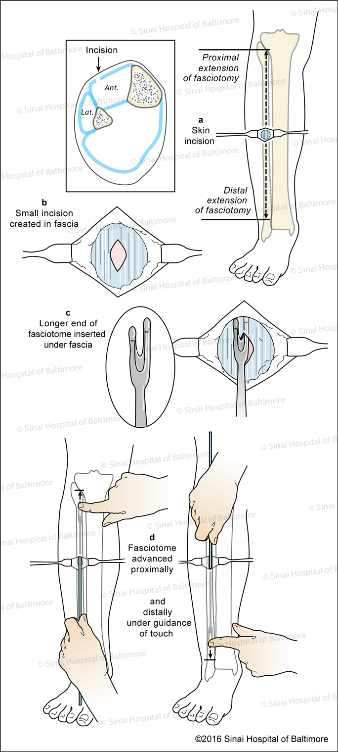
Prophylactic Anterior Compartment Fasciotomy: A, Skin incision; B, Fascial incision; C, Longer end fasciotome inserted under fascia; D, Fasciotome is advanced proximally and distally under guidance of touch.

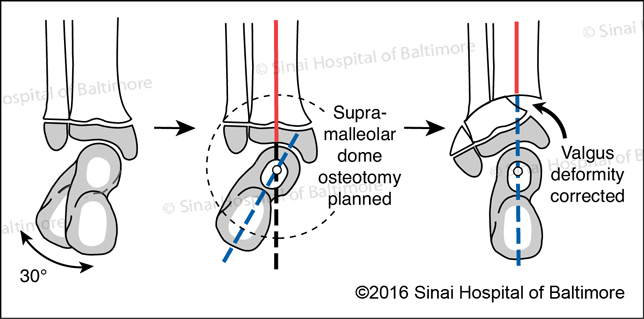
Dynamic Valgus Ankle (Type 2): Supramalleolar Dome Osteotomy Procedure

Bony Realignment of Valgus Ankle for Paley Type 2 Fibular Hemimelia: With a 30 degree valgus deformity a supramalleolar dome osteotomy is planned. After completing the dome osteotomy the valgus deformity is corrected and the ankle and foot are aligned.

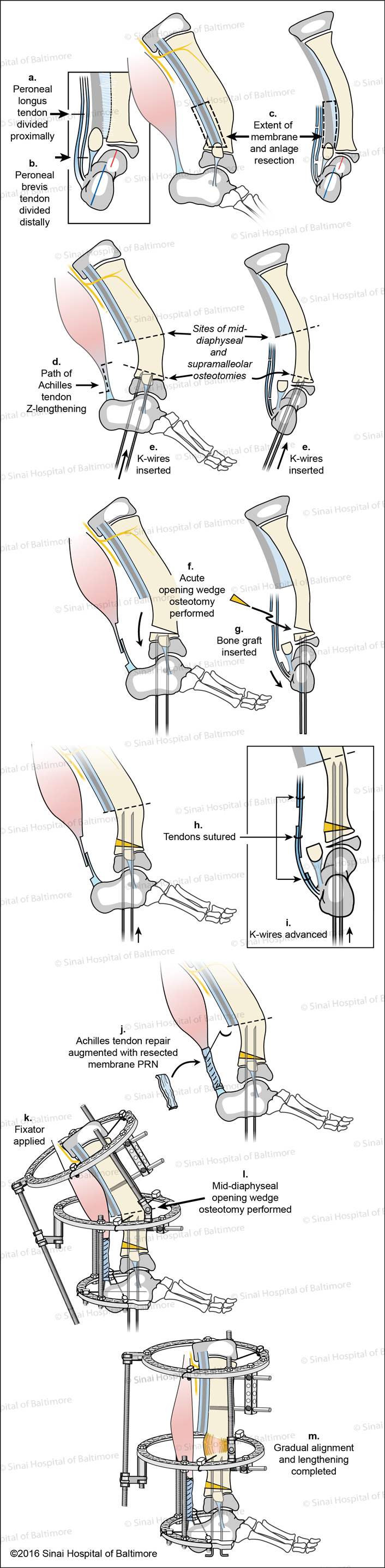
Fixed Equinovalgus: SUPERankle Procedure

Ankle Type (Type 3A) with Supramalleolar Osteotomy

SUPERankle Surgical Technique for Supramalleolar Type Fibular Hemimelia (Paley Type 3A - ankle type ) Fig. A, AP View, Peroneal longus tendon is divided proximally. B, AP View, Peroneal brevis tendon is divided distally. C, Lateral and AP views, The extent of the membrane and anlage resection is identified; D, E, AP and Lateral views, Path of Achilles tendon Z-lengthening, sites of mid-diaphyseal and supramalleolar osteotomies are identified, k-wires are inserted; F, Acute opening wedge osteotomy performed; G, Bone graft is inserted; H, Tendons are sutured; I, K-wires are advanced; J, Achilles tendon repair is augmented with resected membrane as needed; K, Fixator is applied; L, Mid-diaphyseal opening wedge osteotomy is performed; M, Gradual alignment and lengthening is completed.

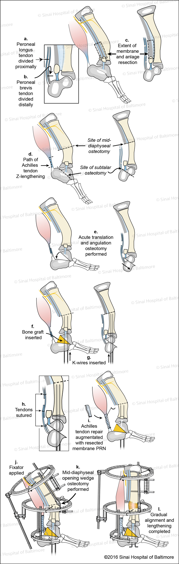
Subtalar Type (Type 3B) with Subtalar Osteotomy

SUPERankle Surgical Technique for Subtalar Type Fibular Hemimelia (Paley Type 3B) Fig. A, Peroneal longus tendon is divided proximally. B, Peroneal brevis tendon is divided distally. C, The extent of the membrane and anlage resection is identified. D, The path of the Achilles tendon Z-lengthening and site of mid-diaphyseal and subtalar osteotomies are identified. E, Acute translation and angulation osteotomy are performed. F, Bone graft is inserted; G, K-wires are inserted; H, Tendons are sutured; I, Achilles tendon repair is augmented with resected membrane as needed; J, The external fixator is applied; K, A mid-diaphyseal opening wedge osteotomy is performed; L, Gradual alignment and lengthening is completed.

Combined Ankle and Subtalar Type (Type 3C) with Supramalleolar and Subtalar Osteotomies

SUPERankle Surgical Technique for Combined Subtalar and Supramalleolar Fibular Hemimelia (Paley Type 3C) Fig. A, Peroneal longus tendon is divided proximally. B, Peroneal brevis tendon is divided distally. C, The extent of the membrane and anlage resection is identified. D, The path of the Achilles tendon Z-lengthening is identified along with the sites of the mid-diaphyseal, supramalleolar, and subtalar osteotomies. E, Acute subtalar osteotomy completed along with translation and angulation. F, Subtalar bone graft is inserted. G, K-wires are inserted. H, Acute supramalleolar wedge osteotomy is performed. I, Supramalleolar bone graft is inserted. J, K-wires are advanced. K, Tendons are sutured. L, The Achilles tendon repair is augmented with the resected membrane as needed. M, The fixator is applied. N, The location of the mid-diaphyseal opening wedge osteotomy is identified. O, Gradual alignment and lengthening is completed.

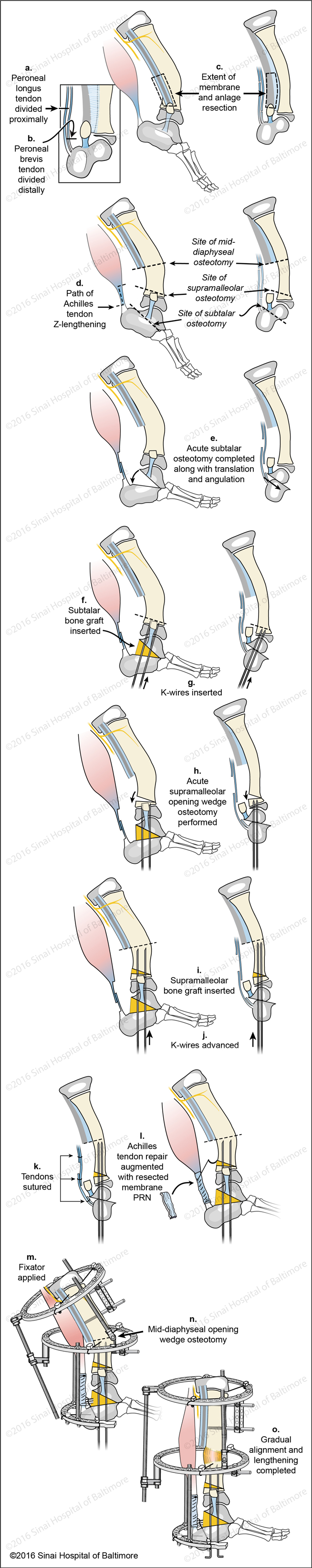
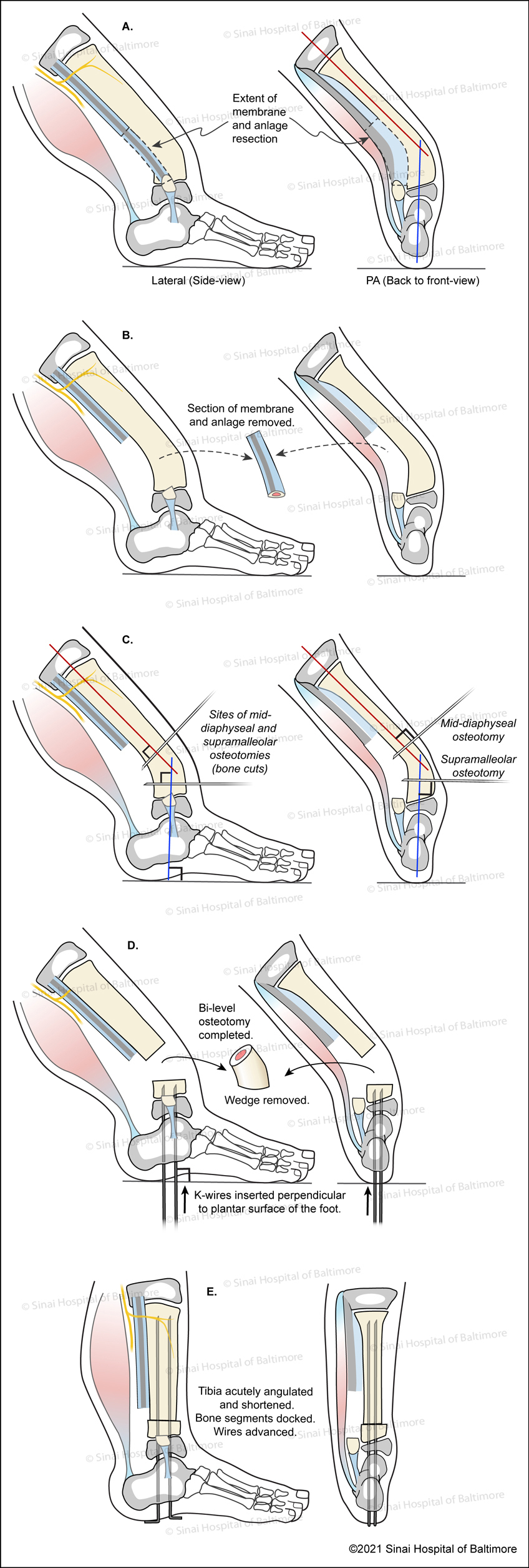
SUPERankle With Shortening (Mid-diaphyseal and Supramalleolar Osteotomies)

SUPERankle Surgical Technique for Supramalleolar Type Fibular Hemimelia (Paley Type 3A - ankle type) Fig. A, AP View, Peroneal longus tendon is divided proximally. B, AP View, Peroneal brevis tendon is divided distally. C, Lateral and AP views, The extent of the membrane and anlage resection is identified; D, E, AP and Lateral views, Path of Achilles tendon Z-lengthening, sites of mid-diaphyseal and supramalleolar osteotomies are identified, k-wires are inserted.