Brachymetatarsia
What is brachymetatarsia?
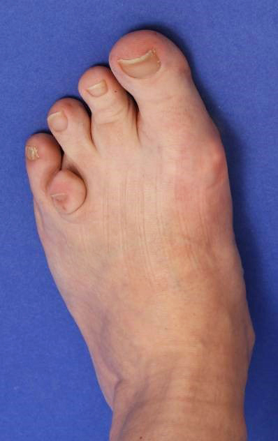
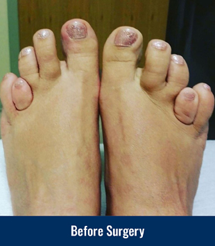
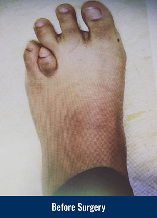
Brachymetatarsia is a condition that can affect the metatarsal bones of the foot. The most common location is the fourth metatarsal. When looking at the foot, the affected toe looks short. This condition can affect either both feet, or just one foot, and it is more common in women than in men.
What are the symptoms of brachymetatarsia?
Symptoms of brachymetatarsia include pain that can occur from walking on the bottom of the feet due to abnormal weight distribution on the front of the foot. Also, the short toe often rides high and rubs on the top of the shoe. Patients can also complain of callus development on the bottom of the foot. Cosmetically, the condition can be a source of embarrassment for certain individuals. Many people with brachymetatarsia avoid going to the pool and beach or wearing flip flops and open sandals.
What is the treatment of brachymetatarsia?
Surgical intervention is designed to lengthen the metatarsal bone to a normal length by means of an osteotomy (cutting of bone) and either gradual or acute lengthening. The surgeon will review the patient’s X-rays to determine how much lengthening is needed. If a patient needs 15 mm (0.59 in) or less, they could be a candidate for acute correction. Those that need more than 15 mm (0.59 in) would do better with gradual correction.
Gradual Lengthening
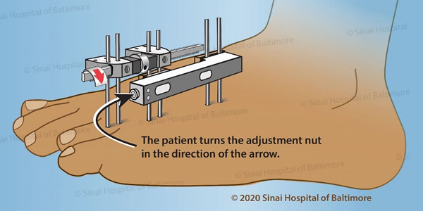 Example of patient instructions to perform gradual lengthening at home
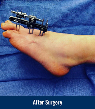
Gradual lengthening involves the use of a miniature external fixator that is surgically applied to the short toe. The patient is able to walk and bear weight immediately after surgery. Once home, the patient performs small adjustments to the external fixator according to the doctor’s instructions. These adjustments will gradually stretch and lengthen the bone and its associated soft tissues until the desired length has been achieved. After lengthening, the external fixator stays on the foot until the bone has fully consolidated.
The time that the external fixator needs to stay on the foot is determined by the rate of lengthening, how much length needs to be gained, and by checking for signs of bone healing on follow-up X-rays. The patient will be lengthening the bone 0.5 mm (0.02 in) per day. Typically after a frame is applied, the patient has to wait for a latency period–the time after surgery that allows for initial healing and preparation for lengthening. The latency period is usually 7-10 days. If a patient needs 15 mm (0.59 in) of gradual lengthening at 0.5 mm (0.02 in) per day, it would take approximately thirty days to achieve normal bone length. The 7-10 day latency period plus the thirty days of lengthening would make it about 37 days for the bone to fully lengthen; however, the bone needs additional time to mature. The initial bone that forms between the lengthened segments is known as the regenerate, and that regenerate product needs to mature into healed bone. This process of consolidation can take another ninety days or so. Therefore, on average, the frame is in place for approximately four months.
The external fixator removal is an outpatient procedure that normally takes place in an operating room. There are some special circumstances when the frame will instead be removed in the office. After the external fixator is removed, the patient is able to walk in protected shoes immediately; they can shower within a few days.
Acute Lengthening
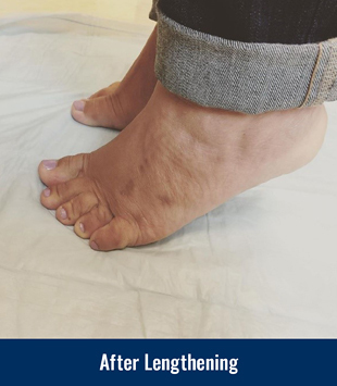
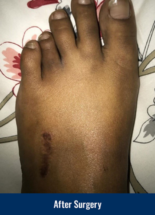
Acute lengthening is a single stage operation that will allow the surgeon to lengthen the bone in one procedure creating a space between the bone ends which is filled with bone graft harvested from the patient’s pelvis or a donor graft, also called an allograft. A low-profile fracture plate is inserted to keep the bone graft in place. After surgery, the patient wears an orthopedic boot for 6–8 weeks and can bear weight on the heel of the foot. After the initial 6–8 weeks, the patient can usually wear their own sneaker. At about 3 months after surgery, the bone is fully consolidated and healed, and the patient can fully weight bear on the foot. The plate does not need to be removed, but can be removed surgically twelve months after the initial surgery.
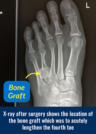
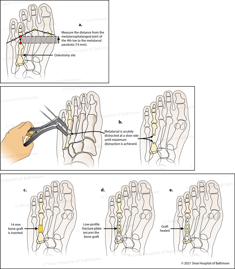 In this graphic depiction of acute surgical treatment for brachymetatarsia of the fourth toe: a. On the patient’s pre-operative X-ray, the surgeon uses established angles of the foot to analyze the length required to normalize the short toe. The bone cut (osteotomy) is planned in the fourth metatarsal bone. b. Surgical instruments are used at the osteotomy site to slowly separate the fourth metatarsal bone until the planned lengthening (distraction) is achieved. c. Next, a bone graft from the patient’s hip is placed into the new metatarsal bone gap. d. and e. A low-profile fracture plate is attached to the bones of the fourth toe to hold the bone graft in place during graft healing.
Why choose the International Center for Limb Lengthening for treatment of brachymetatarsia?
With a collective experience of over thirty years of helping patients with lower leg, foot and ankle problems, the Foot and Ankle Service of the Rubin Institute is one of the leading treatment centers for foot and ankle conditions in the United States. Your doctor will take the time to make sure you understand all of your options and then will customize your treatment to meet your specific needs. Our patients benefit from our team-centered approach with world-renowned surgeons and specialized physician assistants, nurses and physical therapists. We help patients with brachymetatarsia achieve their best possible result.